 |
|---|
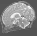
| T2w MRI of newborn |
The CRL is engaged in the development of techniques for generating quantitative biomarkers of disease from newborn brain magnetic resonance imaging (MRI). This is an end-to-end effort beginning with the acquisition of high quality MR images and ending with novel post- acquisition processing methods for extracting statistically useful information from MR scans. Ongoing research focuses on the following areas:
Optimization of clinical and research pulse sequence parameters for newborn imaging at 3T
We are currently acquiring high-resolution T1-weighted and T2- weighted structural images along with high-angular direction (35) diffusion tensor imaging. These sequences have been optimized for the unique size and composition of the smallest newborn brains. Work is ongoing to optimize pulsed arterial spin labeling (PASL) MRI in order to generate a quantitative map of perfusion which can be used to study brain injury related to failures of perfusion.
Additionally, we are investigating techniques for motion-robust MRI to aid in imaging of uncooperative, unsedated neonates. We routinely perform “feed and wrap” imaging of sleeping, unsedated newborns with a high success rate.
Automatic measurement of MRI biomarkers
We have developed a fully-automated, comprehensive analysis pipeline for generating quantitative tissue volumes from newborn MRI. This work includes efforts in post-acquisition artifact correction and noise filtering, inter- and intra-subject registration, the construction of gestational-age specific atlases, the removal of extra-cerebral tissue from MR scans (aka “skull stripping”), optimal statistical classification and related segmentation or image-labeling techniques.
References
- S K Warfield, N I Weisenfeld, C Spencer, and L J Woodward. Prenatal Exposure to Methadone Reduces Brain Tissue Volumes. Presented at a poster symposium at the Pediatric Academic Societies annual meeting in Hawaii, May 1-6, 2008.
- N I Weisenfeld and S K Warfield. Simultaneous Alignment and Central Tendency Estimation for Brain Atlas Construction. Paper read at Statistical Registration: Pair-wise and Group-wise Alignment and Atlas Formation workshop at MICCAI 2007, Brisbane, Australia, Nov 2007.
- N I Weisenfeld, A U Mewes, and S K Warfield. Highly accurate segmentation of brain tissue and subcortical gray matter from newborn MRI. Med Image Comput Comput Assist Interv Int Conf Med Image Comput Comput Assist Interv., 9(Pt 1):199-206, 2006.
- S K Warfield, M Kaus, F A Jolesz, and R Kikinis. Adaptive, template moderated, spatially varying statistical classification. Med Image Anal., 4(1):43-55, March 2000.
- S K Warfield, M Kaus, F A Jolesz, and R Kikinis. Adaptive template moderated spatially varying statistical classification of anatomy. Med Image Comput Comput Assist Interv Int Conf Med Image Comput Comput Assist Interv., Proceedings:431-8, 1998.
- P S Huppi, S K Warfield, R Kikinis, P D Barnes, G P Zientara, F A Jolesz, M K Tsuji, and J J Volpe. Quantitative magnetic resonance imaging of brain development in premature and mature newborns. Ann Neurol., 43(2):224-35, February 1998.
- P S Huppi, S K Warfield, R J Taranto, S A Ringer, F A Jolesz, and J J Volpe. Cortical brain development in preterm and fullterm infants: surface and volume changes measured by 3D-MRI. In: The Society for Pediatric Research Annual Meeting, 41:1733, May 1997. A